Search
Search
Download the Eyetube app to watch 3D videos on your phone.
Alan J. Franklin, MD, PhD
Show Description +
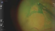
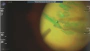
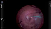
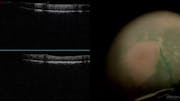
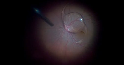
This is the third installment of a three part video series showing repair of a retinal detachment secondary to Stickler Syndrome. The last video showed how to get out of trouble in the posterior portion of the vitrectomy. Here, Alan J. Franklin, MD, PhD, presents a case of a subretinal band that appears as a wrinkle and goes beneath the macular center. The patient's fellow eye is only 20/100. Initially, a drainage retinitomy was performed to permit access under the retina to grasp the subretinal band but the retina was so mobile. A partial perflurorcarbon fill allows the surgeon to perform a peripheral vitrectomy, then the subretinal band is removed with Grieshaber Maxgrip Forceps (Alcon). Part one is available here and Part two is available here.
Posted: 11/27/2017
Alan J. Franklin, MD, PhD
This is the third installment of a three part video series showing repair of a retinal detachment secondary to Stickler Syndrome. The last video showed how to get out of trouble in the posterior portion of the vitrectomy. Here, Alan J. Franklin, MD, PhD, presents a case of a subretinal band that appears as a wrinkle and goes beneath the macular center. The patient's fellow eye is only 20/100. Initially, a drainage retinitomy was performed to permit access under the retina to grasp the subretinal band but the retina was so mobile. A partial perflurorcarbon fill allows the surgeon to perform a peripheral vitrectomy, then the subretinal band is removed with Grieshaber Maxgrip Forceps (Alcon). Part one is available here and Part two is available here.
Posted: 11/27/2017
![Glaukos iStent Inject [3-D]](https://eyetube-thumbs.imgix.net/jgpiy.jpg?auto=compress,format&w=180)


Please log in to leave a comment.